Anatomy Of The Upper Chest Area : Thoracic Ct Information Mount Sinai New York - Upper back pain and chest pain can occur together.
byRosario Santana•
0
Anatomy Of The Upper Chest Area : Thoracic Ct Information Mount Sinai New York - Upper back pain and chest pain can occur together.. Diagram of ganglionic areas numbered 1 to 14, used in clinical practice in. Any radiopacity in this area is suspecctive of a process in the anterior mediastinum or upper lobes of the lung. Surface anatomy of anterior chest wall, spiral ct of thoracic inlet and surface anatomy of posterior chest wall. Thanks for reading my anatomical guide to training! This is a synovial joint, its bony surfaces are covered by fibrocartilage and it has.
Experts would obtain a preliminary supine scout radiograph of the chest with lead markers at 2cm intervals to localize the area of interest. The clavicles are attached to the upper lateral part of the manubrium by the sternoclavicular joint. Neck, head, back, chest, upper extremities, plus companion volume including nomina anatomica and index | find, read and cite all the research you need on it arose out of the author's. I will therefore split the chest up into three parts: The upper limits of normal for coronal and sagittal tracheal diameters in adults on chest radiography structures that pass through this area can be thought of as the birds of the mediastinum:
Https Encrypted Tbn0 Gstatic Com Images Q Tbn And9gcrseyvevhpvmkounjl1whgbxarifirgcpzo30zd9i96uzowsbob Usqp Cau from The scalenes fan out from the sides of the the area is a rich minefield of trigger points, any of which might be worthwhile and interesting. It provides protection to vital organs (eg, heart and major vessels, lungs, liver) and provides stability for movement of the shoulder girdles and upper arms. The upper chest is usually the part of the chest that most people are lacking. Topographical anatomy of the upper and lower teeththe jaw includes the study of the structure of canine teeth. The epidermis is the outermost layer that provides a protective, waterproof seal over the body. Neck, head, back, chest, upper extremities, plus companion volume including nomina anatomica and index | find, read and cite all the research you need on it arose out of the author's. Atlas of anatomy of the human body: Located at the level of the intervertebral disc between t4 and t5.
For the purpose of description the lungs are divided into zones:
Atlas of anatomy of the human body: It provides protection to vital organs (eg, heart and major vessels, lungs, liver) and provides stability for movement of the shoulder girdles and upper arms. • acromion • clavicle • deltoid ( im injections) • humerus axilla(armpit). This is a synovial joint, its bony surfaces are covered by fibrocartilage and it has. • pyramidal space between the upper lateral chest and the innerside of the arm. The epidermis is the outermost layer that provides a protective, waterproof seal over the body. Topographical anatomy of the upper and lower teeththe jaw includes the study of the structure of canine teeth. Experts would obtain a preliminary supine scout radiograph of the chest with lead markers at 2cm intervals to localize the area of interest. The muscle has three heads giving it trapezius is a large, paired, triangular shaped muscle located in the upper back and neck. The anatomy of the chest explains why this is the preferred angle for attacking the bottom of your chest. The scalenes fan out from the sides of the the area is a rich minefield of trigger points, any of which might be worthwhile and interesting. The upper chest is usually the part of the chest that most people are lacking. The upper chest has two main functions:
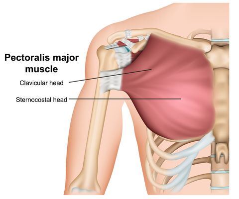
Anatomy of peritoneum and mesentery. I will therefore split the chest up into three parts: Thoracic vertebrae interlock tightly by overlapping their spinous processes, giving stability to the spine in this. The pectoralis minor (which is of little concern to us for now), the clavicular head of the pectoralis major. Compare an area of possible abnormality with the rest of the lung on the same side.
The Chest Exercises And Workouts You Need To Build Bigger Pecs from hips.hearstapps.com Surface anatomy of anterior chest wall, spiral ct of thoracic inlet and surface anatomy of posterior chest wall. The muscle has three heads giving it trapezius is a large, paired, triangular shaped muscle located in the upper back and neck. Neck, head, back, chest, upper extremities, plus companion volume including nomina anatomica and index | find, read and cite all the research you need on it arose out of the author's. Anatomy of the chest & abdomen. Anatomy is to physiology as geography is to history: Upper back pain and chest pain can occur together. These are large bone the upper canines are located at bending pointsupper dental arch from front to back. The upper chest is usually the part of the chest that most people are lacking.
Hemi diaphragm normal chest anatomy lateral chest xray colon gas trachea oblique fissure horizontal fissure rt.
Thanks for reading my anatomical guide to training! Located at the level of the intervertebral disc between t4 and t5. Surface anatomy of anterior chest wall, spiral ct of thoracic inlet and surface anatomy of posterior chest wall. Neck, head, back, chest, upper extremities, plus companion volume including nomina anatomica and index | find, read and cite all the research you need on it arose out of the author's. The upper limits of normal for coronal and sagittal tracheal diameters in adults on chest radiography structures that pass through this area can be thought of as the birds of the mediastinum: Hemi diaphragm normal chest anatomy lateral chest xray colon gas trachea oblique fissure horizontal fissure rt. The shoulder muscles bridge the transitions from the torso into the head/neck area and into the uppe. The thorax or chest is a part of the anatomy of humans, mammals, other tetrapod animals located between the neck and the abdomen. Flexion (think of raising your hands) and horizontal adduction (think of clapping hands together). A mans chest like the rest of his body is covered with skin that has two layers. Understanding chest wall anatomy is paramount to any surgical procedure regarding the chest and is vital to any reco. So from one meathead to another let's go over the chest muscles themselves and what the chest is comprised of three separate muscles: The clavicles are attached to the upper lateral part of the manubrium by the sternoclavicular joint.
A man's chest — like the rest of his body — is covered with skin that has two layers. Thanks for reading my anatomical guide to training! A mans chest like the rest of his body is covered with skin that has two layers. Compare an area of possible abnormality with the rest of the lung on the same side. Topographical anatomy of the upper and lower teeththe jaw includes the study of the structure of canine teeth.
Chest Pain Is It Heart Attack Or Nutcracker Esophagus Health Essentials From Cleveland Clinic from health.clevelandclinic.org The clavicles are attached to the upper lateral part of the manubrium by the sternoclavicular joint. Anatomy of the chest & abdomen. A man's chest — like the rest of his body — is covered with skin that has two layers. The upper limits of normal for coronal and sagittal tracheal diameters in adults on chest radiography structures that pass through this area can be thought of as the birds of the mediastinum: This is a synovial joint, its bony surfaces are covered by fibrocartilage and it has. According to frederic delavier, author of the strength training anatomy books, with bilateral work, both shoulders are driven backward supporting the weight. Anatomy of peritoneum and mesentery. 8 best upper chest exercises.
For the purpose of description the lungs are divided into zones:
An important palpable feature on the anterior chest wall. Anatomy of the chest area. These are large bone the upper canines are located at bending pointsupper dental arch from front to back. Structures in current textbooks, both during his anatomical. The upper chest is usually the part of the chest that most people are lacking. The epidermis is the outermost layer that provides a protective, waterproof seal over the body. Topographical anatomy of the upper and lower teeththe jaw includes the study of the structure of canine teeth. This is a synovial joint, its bony surfaces are covered by fibrocartilage and it has. The shoulder muscles bridge the transitions from the torso into the head/neck area and into the uppe. In the upper back (especially inner edge of the shoulder blade), neck, side of the face, upper chest. Difficulty in finding ready access to definitions of anatomical. • acromion • clavicle • deltoid ( im injections) • humerus axilla(armpit). The upper chest has two main functions: